Fascinating images of creatures, substances and structures at a scale so tiny that they normally go unseen by human eyes.
Nikon just announced the winners of the 2017 Small World Photomicrography Competition, and below are some of the winning and honored images. The contest invites photographers and scientists to submit images of all things visible under a microscope. More than 2,000 entries were received from 88 countries in 2017, the 43rd year of the competition.
(Source: The Atlantic)
Nikon just announced the winners of the 2017 Small World Photomicrography Competition, and below are some of the winning and honored images. The contest invites photographers and scientists to submit images of all things visible under a microscope. More than 2,000 entries were received from 88 countries in 2017, the 43rd year of the competition.
 |
| The 12th place winner shows a closeup of the eye of an Opiliones (daddy longlegs) magnified 20x.Charles Krebs, Issaquah, Washington |
 |
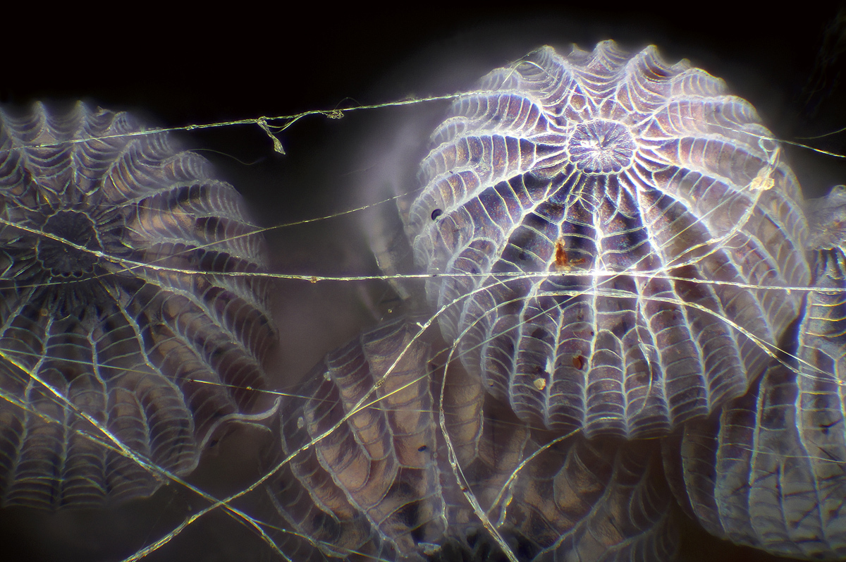
| 2nd Place: A Senecio vulgaris (a flowering plant) seed head, magnified 2x.Dr. Havi Sarfaty, Yahud-Monoson, Israel |
 |
| 11th place shows an image of plastic fracturing on a credit card hologram magnified 10x.Steven Simon, Grand Prairie, Texas |
 |
| Image of Distinction: Nsutite and Cacoxenite (minerals), magnified 5x.Emilio Carabajal Márquez, Madrid, Spain |
 |
| Honorable Mention: The eyes of a jumping spider, magnified 6x.Emre Can Alagöz, Istanbul, Turkey |
 |
| The 15th place winner is an image of a 3rd trimester fetus of a Megachiroptera (fruit bat), magnified 18x. Dr. Rick Adams, Greeley, Colorado |
 |
| Image of Distinction: Moth eggs in spider silk, magnified 16x. Walter Piorkowski, South Beloit, Illinois |
 |
| Image of Distinction: A natural bridge (petiole nodes) connecting the abdomen and thorax of an ant, magnified 5x.Can Tunçer, Izmir, Turkey |
 |
| 8th Place: An image of a newborn rat cochlea with sensory hair cells (green) and spiral ganglion neurons (red), magnified 100x.Dr. Michael Perny, Bern, Switzerland |
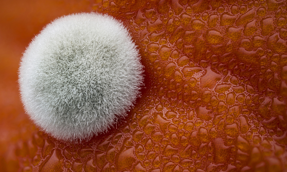 |
| 5th Place: Mold on a tomato magnified 3.9x.Dean Lerman, Netanya, Israel |
 |
| Honorable Mention: A Taraxacum officinale (dandelion) cross section showing curved stigma with pollen, magnified 25x.Dr. Robert Markus, Nottingham, United Kingdom |
 |
| Image of Distinction: Simple eyes of an Ectemnius (digger wasp), with condensation, magnified 20x.Laurie Knight, Tonbridge, United Kingdom |
 |
| The 7th place winner shows individually labeled axons in an embryonic chick ciliary ganglion magnified 30x.Dr. Ryo Egawa, Nagoya, Japan |
 |
| Image of Distinction: A Cladocera (water flea) magnified 10x.Rogelio Moreno, Panama City, Panama |
 |
| Image of Distinction: A group of rotifers magnified 20x.Frank Fox, Konz, Germany |
 |
| Image of Distinction: The face of a small moth magnified 5x.Jan Rosenboom, Rostock, Germany |
 | |
| 9th Place: Growing cartilage-like tissue in the lab using bone stem cells (collagen fibers in green and fat deposits in red), magnified 20x for collagen; 40x for fat deposits.Catarina Moura, Dr. Sumeet Mahajan, Dr. Richard Oreffo & Dr. Rahul Tare, Southampton, United Kingdom |
 |
| Image of Distinction: Abdominal proleg of a Lasiocampa (caterpillar) magnified 3.7x.Dean Lerman, Netanya, Israel |
 |
| The 4th place winner shows the everted scolex (head) of a Taenia solium (tapeworm), magnified 200x.Teresa Zgoda, Rochester, New York |
 |
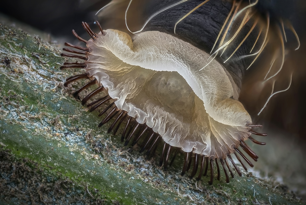
| 18th place shows Synapta (sea-cucumber) skin magnified 100x.Christian Gautier, Le Mans, France |
 |
| Honorable Mention: Warp knitted curtain fabric magnified 10x.Marc Van Hove, Zwijnaarde, Belgium |
 |
| Honorable Mention: Liquid crystal magnified 500x.Michael Tuchband, Boulder, Colorado |
 |
| Image of Distinction: Mosquito larva, early development, magnified 10x. Charles Krebs, Issaquah, Washington |
No comments:
Post a Comment